Movement is the act of changing position/ posture by the whole organism or part of the organism
Types of movement
1. Movement of curvature (growth movement)
2. Movement of locomotion
1. MOVEMENT OF LOCOMOTION
This is the type of movement where by the whole organism moves from one place to another
Movement in locomotion is shown in all animals and some protoctists exhibit variety of movements. Animals and some protoctists exhibit variety of movements, these are
- Amoeboic
- Ciliary
- Muscular
-
Flagella
edu.uptymez.com
I. AMOEBA MOVEMENT
Is the type of movement exhibited by some protozoans such as Amoeba and white blood cell (WBC); amoeba movement is caused by streaming of the cytoplasm towards a peripheral region of the cell resulting into projections known as PSEUDOPODIUM
The cytoplasm steaming into these projections is withdrawn from others and flows in one direction to bring about movementCILIARY
II. MOVEMENT
This is the type of movement where by some (protozoan) organism use cilia for movement
These protozoans are paramecium and larvae of some aquatic animals. The body of such organism is covered by thousands of small hair like structures called cilia. Movement is brought about by word noted backward and forward beating of cilia. The backward pushing of water (propels) pushes the organism forward
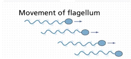
III. FLAGELLAR MOVEMENT
This is the type of movement exhibited by some organisms which possess flagella.
Such organisms include Euglena, chlamydomonas, trypanosome and some bacteria.
Flagella are very similar in structure to cilia but are much longer than in euglena, the whipping of the flagellum cause the swirling of the water around the organism. This swirling makes the organism rotate at the same time move forward
IV. MUSCULAR MOVEMENT
This is the type of movement exhibited by the contraction and relaxation of muscles. Since muscles alone cannot bring about fast movement, most animals have a firm and hard base for support and attachment of muscle. This firm and hard base is called skeleton.
Importance of movement in animal and plant
- Organism moves in search of food and shelter
edu.uptymez.com
• Organism move away from a negative stimulus, e.g. predator, chemical, fires, to secure protection.
• Movement enables animal to come together for mating
• Movement enables organism to move towards the positive stimulus for instance growth factor such as light, gravity and water.
MOVEMENT OF THE HUMAN BODY
Contraction and relaxation of muscles cause muscular movement in vertebrates animals such as man.
Movement of the human body is made possible by supportive structure like skeleton which provides attachment of muscles and other body organs. The body is supported by skeleton. The muscle fibres become shorter on contraction. Muscles are paired producing movement in opposite direction.
One muscle contracts while the other is relaxed, this is called antagonistic
action
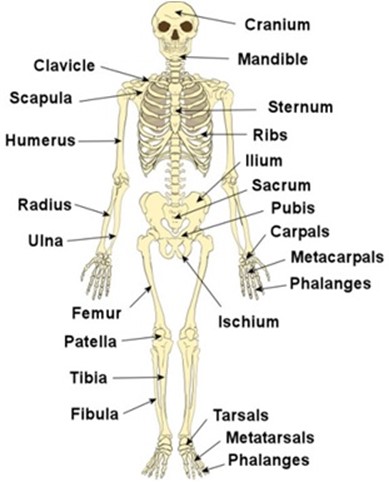
THE HUMAN SKELETON
Skeleton is a frame work of tissue supporting a human or animal’s body
The human/ mammalian skeleton consist of the following major parts
1) Skull
2) Vertebral column
3) Limb
4) Girdles
The human skeleton is made up of separate units which are joined together; the points of junctions where 2 units meet are called joints
The skull sternum,ribs and the vertebral column form the axial skeleton. The limbs and limbs girdles form appendicular skeleton
THE HUMAN SKELETON
TYPES OF SKELETON
There are 3 types of skeleton
I. Hydrostatic skeleton
II. Exoskeleton
III. Endoskeleton
I. HYDROSTATIC SKELETON
This is a skeleton found in soft bodied animals. The body tube is filled with fluid that produce pressure when muscles around it contract bring about movement e.g. Earthworm
II. EXOSKELETON
These are skeleton found outside of the body which is typical arthropods e.g. insect
III. ENDOSKELETON
This is a raid frame work of bones cartilages surrounded by muscles that contract and relax bringing about a movement.
Bone – is the one of the hardest tissue and found only in vertebrate.
Cartilage – is softer and more flexible tissue than bones. In animal cartilage found in nose, part of ear and on the end of bones
FUNCTIONS OF SKELETON
1. Support
The skeleton provides a rigid frame work which supports softer parts of the body. (Provides attachment for muscles and body organs)
2. Locomotion
The skeleton enables the organism to move from one place to another.
3. Protection
It protects delicate internal organs. Example the skull protects the brain. The sternum protects spinal cord and ribcage protects the lungs and heart.
4. Formation of blood cells
Red blood cells and white blood cell are made/ manufactured in the bone marrow.
5. Shape
The skeleton gives animals a definite shape
6. It stores minerals such as calcium and phosphorus
The human skeleton system is divided into two major parts
1. The Axial skeleton
2. The Appendicular skeleton.
1. THE AXIAL SKELETON
The axial skeleton consist of four parts which are
- The skull
- Ribcage
- Vertebral
- Sternum
edu.uptymez.com
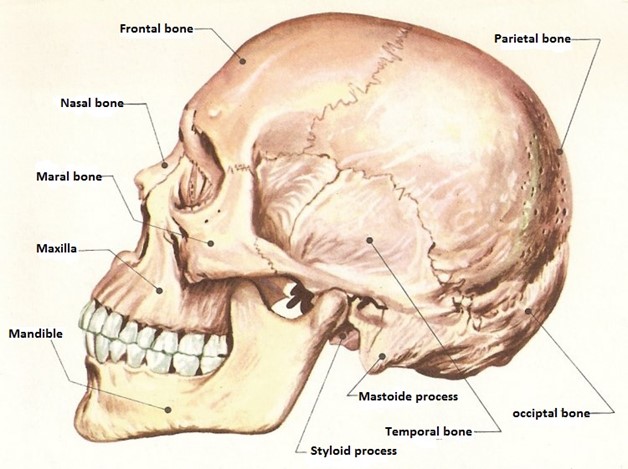
The skull
Is made up of small bones joined together to form the cranium. The bones are joined together by irregular edges called sutures which are immovable joints
- It acts like a box enclosing and protecting the brain, parts of the inner ear, nose and eyes.
- It consists of the upper and lower jaw / bones which hold teeth
- Parts of the skull form hollows which protect the eyes (orbits) and ears
-
The main function of the skull is to protect the brain, olfactory organs, middle and inner ear and the eyes.
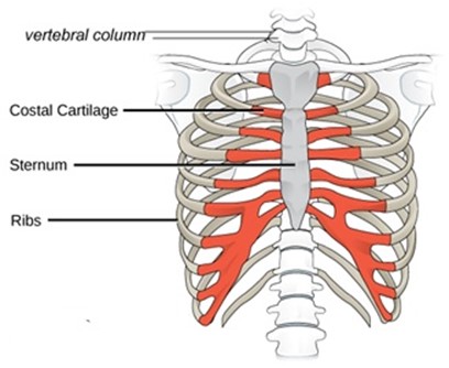
- Ribcage and sternum
edu.uptymez.com
edu.uptymez.com
Skull is composed of bones of the sternum and the ribs. These bones form a thoracic cage which encloses the thoracic cavity, protecting heart, lungs and major blood vessels.
It consists of 12 pairs of ribs joined to thoracic vertebrae at the back and sternum at the front.
The last 2 ribs are not joined at the sternum are known as floating ribs.
This arrangement enables a protective cage bones to be formed which enclose the heart and lung. Between the ribs are intercostals muscles. The ribs are associated with the axial skeleton
The sternum consists of small bones known as Sternebrae. The sternum forms part of the ribcage and provides surface for attachment of ribs
III.
Vertebral column
This is the main axis of the body.
It is made up of small bones (33) known as Vertebrae. Between two adjacent vertebrae is a cartilage known as intervertebrate disk which act as shock absorbers, and reduce friction
The main function of the vertebral column is to support the body and support the spinal cord. The backbones have five types of vertebrae, which are:
a) Cervical
b) Thoracic
c) Lumber
d) Sacral
e) Caudal
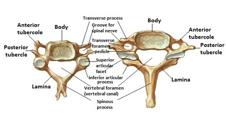
a) Cervical vertebrae
There are 7 short cervical vertebrae, found in the neck region. The first is below the skull is atlas followed by the axis.
Atlas articulates with the skull to allow nodding movement of head.
The axis allows rotational movement of the atlas which acts as a pivot. This allows turning / side to sideways movement of the head. (Shake the head to say no), also cervical vertebrae support the head region and protect blood vessels that pass through their canals. They also provide surface for the attachment of the neck muscles
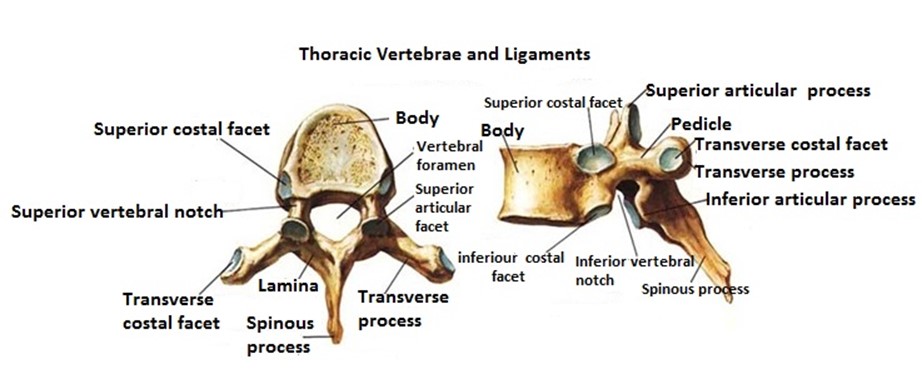
b) Thoracic vertebrae
Are found in the chest region, they are 12 vertebrae. The thoracic vertebrae with the ribs and sternum form the thoracic cage.
The main role of the thoracic cage is to protect the heart, lungs and major blood vessels also plays major role on breath movement
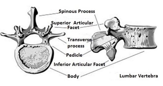
c) Lumbar vertebrae
There are five (5) lumbar vertebrae in human, seven in rabbits and six (6) in rats
They are short bones found in the abdominal region. Lumbar vertebrae have a number of projections that provide surface for attachment of abdominal muscles and muscles of the lower half of the back. The large thick Centrum gives support to the upper half of the body.
Lumbar vertebrae permit bending, sideways movement and rotation of the trunk. This is the region where large muscles of the abdomen are attached
d) Sacral vertebrae
Sacral vertebral are focused together to form the sacrum, they are found in the sacral region. Sacrum provides a large surface area of the attachment of muscles of the back.
e) Caudal vertebrae
These are found in the tail region. The number of caudal vertebrae varies from one animal to another depending on the size of the tail. In man there is no external tail, there are four caudal vertebrae which are used to form which is (no functions)
2) THE APPENDICULAR SKELETON
The appendicular skeleton is composed of the appendage limbs which are attached to the axial skeleton
There are 2 types of limbs namely
- Fore limbs
- Hind limbs
edu.uptymez.com
I. FORELIMBS
Forelimbs are attached to the axial skeleton to the anterior part of the body. Forelimbs comprise the following parts
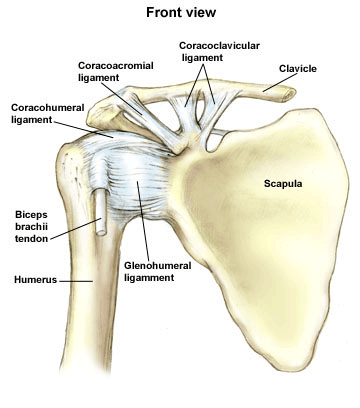
Pectoral girdle
b) Humerus – Is long bone of the upper arm and provide surface for attachment of muscle
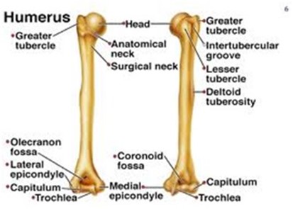
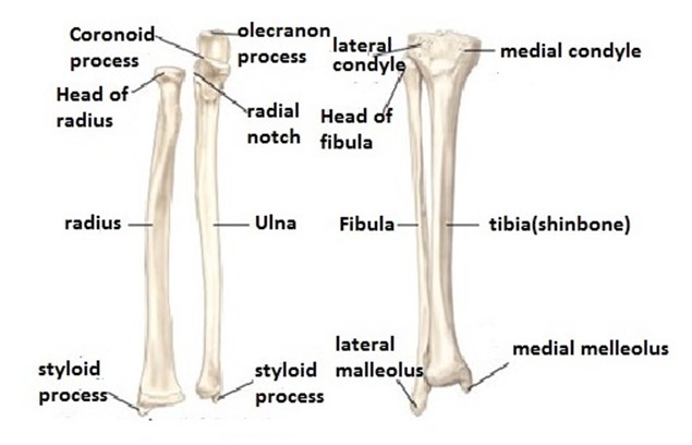
c) Ulna and Radius
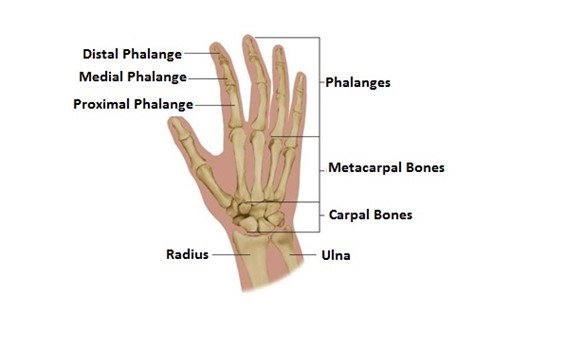
d) Carpals, metacarpals and phalanges
– Carpals are nine small bones which form the wrist. They articulate with radius and ulna at the upper and metacarpus at the lower ends
i) They allow free movement of hands and wrist.
ii) They provide surface for attachment of wrist muscles
Metacarpals are five slightly elongated bones which are found in the palm
-
Each of them articulate with phalange of finger bone
1) They provide surface of attachments of palms muscles
2) They support and maintain shape of the arm.
Phalanges
edu.uptymez.com
Phalanges form the skeleton of the fingers
- HIND LIMBS
edu.uptymez.com
Hind limbs are attached to the axial skeleton to the posterior part of the body. Hind limbs comprise of the following
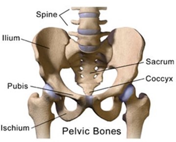
a) Pelvic girdle
Is made up of several bones found around the hip region; It contains 2 halves, the left and right. Each half lies on either side of the vertebral column. In this way it supports the hind limbs.
Pelvic girdles have two bones known as pubic bones, each pubic bone comprises of three (3) bones known as ischium, ilum and pubis. The ischium and ilium are fused together
The size of the pubic cavity is very important in females during birth. Causing the widening of the female girdle.
- The pelvic girdle forms a protective cage around vital organs such as female reproductive organs.
- It also supports legs, articulating with the head of femur to form hip joint.
- It articulates with sacrum and provides for a tail where it is present.
edu.uptymez.com
b) Femur
Is a long bone on the upper part of the hind limb (on the thigh region)
- The head of femur fits in the pelvic girdle to form hip joint
- It articulates with tibia at lower end to form knee joint
- It provides surface for the attachment of leg muscles and it supports the thigh.
edu.uptymez.com
c) Tibia and fibula
These are long bones of the lower
- Tibia is a very long bone, found on the side of the big toe. It may be free or partly fused to the smaller fibula which lies alongside it.
-
Fibula is much smaller in size and fused to the tibia in the lower part of the leg.
A small round bone is called patella/ knee cap lies in front of the knee joint, it prevents the leg from bending up wards at the knee.
- The tibia and fibula supports the front part of the leg below knee
- They provide surface for attachment of the knee (shin) muscles.
- They articulate with femur to form knee joint, and with metatarsals to form the ankle joint.
-
Red blood cells are manufactured in the tibia and fibula bone marrow.
edu.uptymez.com
d) Tarsals, metatarsals and phalanges
– Tarsals are six (6) small bones in the ankle. Two of them are elongated and one projects backwards to form a heel bone. The tarsals provide surface for attachment of ankle muscle. The heel bone prevents the foot from bending backwards.
– Metatarsals
These are elongated bones in foot.
There are 5 in humans and in most animals. Each one leads to a phalange. The metatarsals provide surface for attachments of foot muscles, they also support and maintain the shape of the foot.
Functions
- Tarsals articulate with fibula to form the ankle joint
- Tarsals articulate with metatarsals to form the foot
-
Metatarsals articulate with phalanges to form toes
SKELETON OF HUMAN FORE LIMB