MEIOSIS:
Meiosis is the type of nuclear division which result with four daughters cucle each havinghalf the number of chromosomes of the parent cell. It is also termed as REDCTION DIVISION as it reduces the number of chromosomes from diploid (2n) to haploid (n). it is mainly a means of gametes formation …………………….
The human gamete has 23 chromosomes, 33 of which are autosomes and 1 sex chromosome of sparm cell has 23 chromosome
i.e. 22A + 1 sex chromosome
22A + x or y sex chromosome
An ovam:
22A + 1 sex chromosome
22A + x chromosome
SOME TERMS USED:
- CHROMOSOME: A thread like structure visible in the nucleus of a cell during nuclear division
- SEX CHROMOSOE: Chromosomes responsible for determination of sex of an individual
- AUTOSOME: Chromosome responsible for determination of characters other than sex
edu.uptymez.com
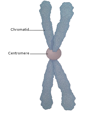
Fig .Structure of Chromosome
-
PHASES OF MEIOSIS:
Meiosis is a long process as it passes through two cycles to completion
(a) Meiosis I or first meiotic division
(b) Meiosis II or second meiotic division
- MEIOSIS I
edu.uptymez.com
This reduces the number of chromosomes to half.
Meiosis I has the following phases:
(i) Interphase I
(ii) Prophase I
(iii) Metaphase I
(iv) Anaphase I
(v) Telophase I
(i) INTERPHASE I
It is a preparatory phase during which the nucleus is about to start dividing. The events of interphases one include the following:
(a) Replication of organalles
(b) Increase in size of the cell
(c) Replication of most of DNA and histories
(d) The chromosomes replicate so that each of them exist as a pair of chromotids being joined together by the centromere
(e) The chromosomal material will but no structure is clearly visible except the nucleoli
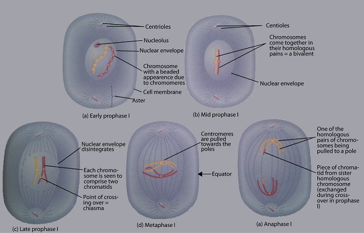
(ii) POOPHASE I
This is the largest of all stages. It is often described in five stages consecutive stages namely:
(a) Laptotene
(b) Zygotene
(c) Pachytene (lezypadid)
(d) Diplotene
(e) Diakinesis
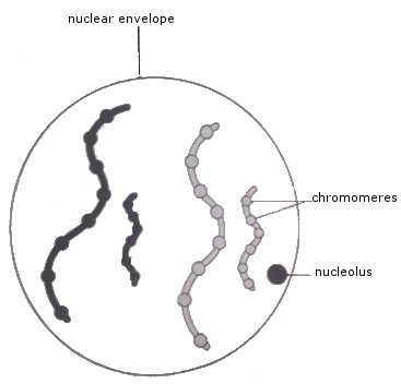
(a) LEPTOTENE (thin tread stage)
Leptetene stage initiates meiosis. During this stage:
(i) Chromosomes appear as unioiled thread like
(ii) Chromosomes appear to be longitudinally single
(iii) Chromosomes appear to have dense granules which occur at irregular intervals along their lengths. These are called choromosomeres
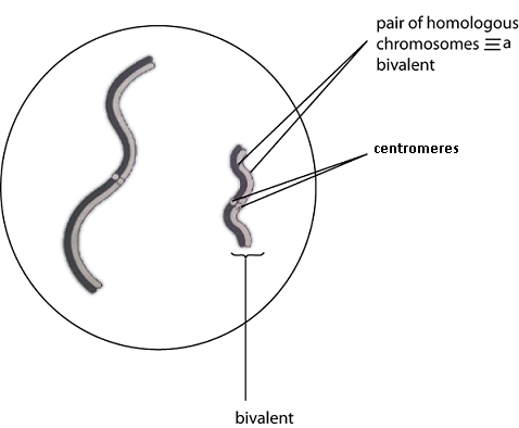
(b) ZYGOTENE (pairing stage)
This is initiated by the movement of chromones in the zygotene stage.
(i) Homologous chromosomes more close to one another and up they lie side by side, chromosomere by chromosomere under the influence of attraction force called SYNAPTIC FORCE
(ii) Synapsis begins at one or more points along the chromosome and unites along the entire length
(c) PACHYTENE (Thickening stage)
During pachytene
(i) Chromosomes are thickened and shorted by coiling and become visible
(ii) The nucleolus is attached to particular chromosomes
(iii) The synaptic force of attraction start to lapse and homologous chromosomes start to separate from each other.
Each chromosome appears a double structure.
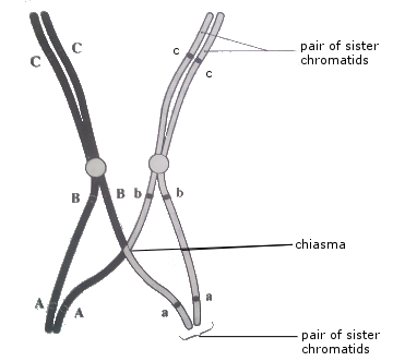
(d) DIPLOTENE (Duplication stage)
(i) There is a complete duplication of each chromosome to produce two chromatids, thus each bivalent has four chromatids
(ii) The chromatids of homologous chromosomes cross over one another. At the point called chiasmata or cros over the number of chiasmata to be formed depends on the length of the chromosome. At the chiasmata, chromosomes breaks and rejoin, thereby exchanging hereditary materials. As a result, genes from materinal chromosomes exchange with genes from paternal chromosomes leading to new gone combination in the resulting chromatids. This is a means of bringing about variation
edu.uptymez.com
(e) DIAKINESIS (Moving apart stages)
During Diakinesis:
(i) The nucleolus detaches from its special bivalent and disappears.
(ii) The chiasmata tend to lose their original position and move towards the ends of chromosomes
(iii) The bivalent become considerably more contructed
(iv) Chromotids if homologous chromosomes continue to repel
(v) The contriolas of present migrate to the poles
(vi) The nuclear membrane starts to disintegrate and spindle fibre start to form
Study problem:
-
Describe the events of prophase of meiosis I and comment on biological consequences of chiasmata formation.
MATAPHASE I
During Metaphase of meiosis
(i) The bivalent are arranged across the equitorial plate of the spindle with each centromere equidistant from the equatorial plate
(ii) The nuclear membrane has broken down completely
(iii) The spindle fibre forms and hold the centromers at the equator
ANAPHASE I
During this phase:
(i) The two centromenes of each bivalent do not divide, instead the siste chromatids separate
(ii) The centomere pairs move toward the opposite poles
(iii) The chiasmata contents completely breakdown
(iv) The chromosome are separate into two haploid sets of chromosomes in the daughter cells.
TELOPHASE I
This marks the end of meiosis. During this phase,
(i) The homologous chromosomes arrive at the opposite poles
(ii) Spindle fibre disappears, chromatids uncoil and the nuclear membrane rejoin around each pole
(iii) Cytoplasm dividing to form two daughter cells
N.B: In many plant cells there is no telephase, cell wall formation on interphase I. the cell pass straight from anaphase to prophase II.
INTERPHASE II
This occurs in animal cell only is there is no interphase II in plant cell. Replication of DNA doe snot occur and energy stores of the cell increases. This stage is followed by meiosis II. The behavior of chromosomes in meiosis II is the same as that in meiosis I.
PROPHASE II
During this phase:
(i) Nucleoli and nuclear membrane start to disintegrate
(ii) The chromatids shorter and thicken
(iii) Cantoriols if present move to opposite poles the cell
(iv) Spindle fibre appear
METAPHANE II
The chentromeres align at the equator of the spindle
ANAPHASE II
(i) The centromeres divide
(ii) The spindle fibres shorters and pull the centromeres to opposite poles
(iii) The eytoplasm started to cleave
TELOPHASE II
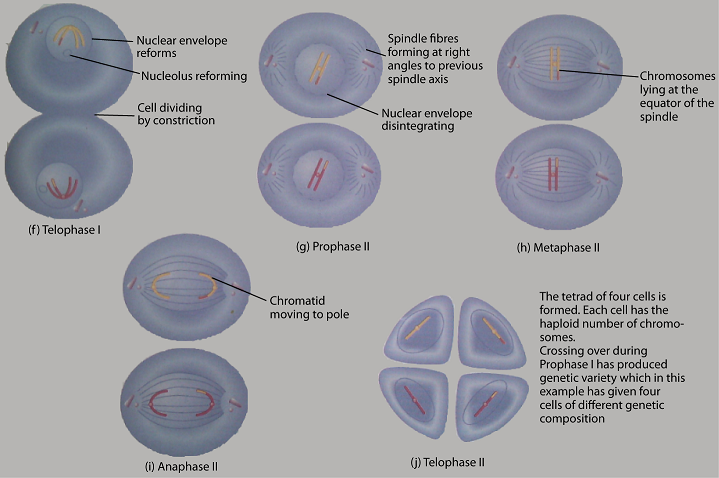
– The chromosomes uncoil
– Spindle fibre disappear
– nuclear membrane reforms followed by complete cytokinesis
– Four daughter cells are formed each with half the number of chromosomes of the parent cell
SIGNIFICANCE OF MEIOSIS:
It insures constant chromosome number to all species which reproduce sexually. This is because during gametes formation. The number of chromosomes is reduced to half and restored at fertilization.
(ii) It provide opportunities for new gene combination through chiasmata formation. Hence a mechanism of variation.
DIFFERENCE BETWEEN MEIOSIS AND MITOSIS
edu.uptymez.com
| STAGE | MITOSIS | MEIOSIS |
| Prophase | – chromosomes not visible – Homologous chromosome remain separate – No chiasmata formation |
– chromosomes visible – Homologous chromosomes pair up – chiasmata formation take place |
| Metaphase | – chromaitd pairs line up on the equator of the spindle centromores line up on the equator | – this occur in metaphase II but no metaphase I – centromere line equidislent aove and below the equater of the spindle |
| Anaphase | -chromatids separate number of chromosome present as parent cell – Separated chromatids identical |
– chromosomes separate in Anaphase I, chromatids separate in Anaphase II – The separated chromosomes and chromatids may not be identical – Half the number of chromosomes is present in daughter cell |
| Telophase | – Both homologous chromosomes are in each daughter cell | – only one of each pair of homologous chromosome is in each daughter cell |
| Occurance | – occurs in the formation of somatu cells | – Occurs in the formation of gametes and spores |
edu.uptymez.com
STAGES OF SEXUAL REPRODUCTION
Sexual reproduction involves the following stages:-
1) gametogenesis
2) Copulation
3) Fertilization
4) Cleavage
5) Implantation
6) Pregnancy
7) Parturition(birth)
8) Parental case.
-
GAMETOGENESIS.
Definition:
Gametogenesis is the general process of gametes formation in both male and female reproducing sexually.
edu.uptymez.com
- Meiosis is the process by which gametes are formed can also be called gametogenesis literally ‘creation of gametes’.
- The type of meiosis in male organism forms a spermatogonium to a primary spermatocyte a secondary spermatocyte a spermatid and finally a spermatozoid is spermatogenesis.
edu.uptymez.com
Definition:
Oogenesis is the process of meiosis in female organism from oogonium to a primary oocyte, a secondary oocyte and then an ovum (egg cell).
- The primordial germ cells once they have been populated the gonalds proliferate into sperm (in testes) or ova (in the ovary).
edu.uptymez.com
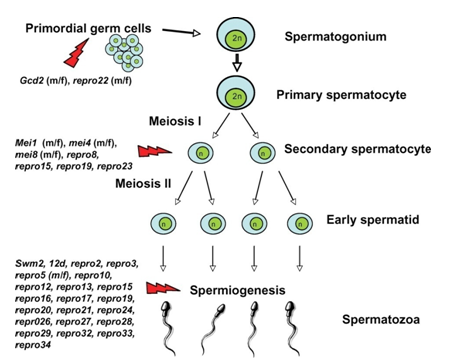
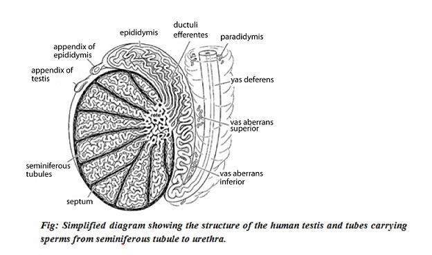
SPERMATOGENESIS
- In male testis there are tiny tubules (seminiferous tubules) containing diploid cells called spermatogonia that develop into mature spermatozoa (spermatozoa are the mature male gametes in many sexually reproducing organisms).
- In spermatogenesis i.e. a process during which spermatogonia (sperm cells) multiply giving rise to other spermatogonia restoring their population and to other which mature to spermatocyte.
-
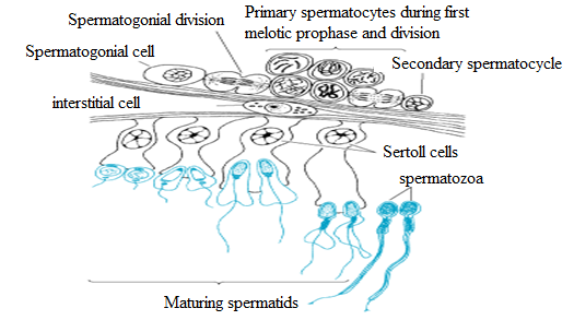
Around the periphery of the seminiferous tubules are located specialized cells known as spermatogonia.
Spermatogonia are diploid cells set aside early in embryonic development. They may divide by mitosis to generate more spermatogonia or by meiosis to produce spermatids each of which will differentiate into a mature sperm cell.
- Spermatogonia destined to undergo meiosis first differentiate into primary spermatocytes which undergo two successive meiosis divisions.
-
After meiosis I the produced cells are called secondary spermatocyte which each in turn undergoes the secondary division become spermatids each containing a unique set of 23 single chromosomes that ultimately mature into four sperm cells (spermatozoa).
- The seminiferous tubular contain two types of cell;
edu.uptymez.com
- Germ cells; these undergo the two division of meiosis to form the spermatozoa
-
Sertoli Cells: Acts as nerve cells ensuring that the germ cells have adequate nourishment.
edu.uptymez.com
Fig: The stages of spermartozoa formation.
- Spermatids undergo transformation in order to become spermatozoa
edu.uptymez.com
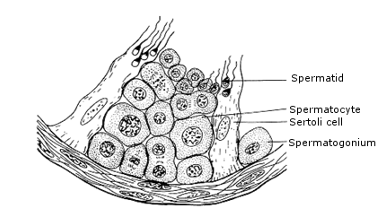
Fig:
Diagram showing the structure of part of the wall of seminiferous tubule.
SPERMATOGENESIS
- Occurs in seminiferous tubules.
- Stored in epididymis.
edu.uptymez.com
Process:
- Diploid spermatogonia divide by mitosis from germinal epithelium (germinal epithelial cells).
- Some of them grow to produce diploid primary spermatocytes.
- Diploid spermatocytes undergo first meiotic division to form two haploid secondary spermatocytes.
- Haploid secondary spermatocytes undergo second meiotic division to form haploid spermatids.
- These grow in shape and become spermatozoan.
- The sertoli cells provide nutrition and protection against body immune system.
edu.uptymez.com
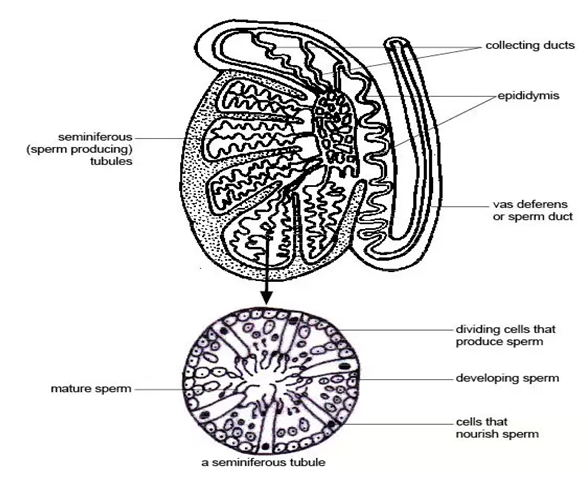
From the figure
The interior of the testis, site of spermatogenesis within the seminiferous tubules of the testis cells called spermatogonia develop into sperm, passing through spermatocyte and spermatid stages. Each
sperm passes as a long tail coupled to a head which contain a haploid nucleus.
MECHANISM OF SPERMATOGENESIS
The process of spermatogenesis is divided into the following phases (as shown below):-
1. Multiplication phase.
2. Growth phase.
3. Maturation phase.
4. Metamorphosis.
1. MULTIPLICATION PHASE
- Also known as spermatocytogenesis.
- Here the sperm mother cells present in the germinal epithelium of the seminiferous tubules divide repeatedly by mitosis to form a large number of diploid rounded sperm mother cells called spermatogonia.
edu.uptymez.com
Some of these sex cells move towards the lumen of seminiferous tubules and enter growth phase. These cells are called primary spermatocytes. The primary spermatocytes are diploid and contain (44 + XY)
chromosomes.
Some of these cells produced by the division of spematogonia remain in the original condition and continues to divide giving rise to primary spermatocytes such cells are known as stem cells.
- GROWTH PHASE
edu.uptymez.com
During this phase, spermatocyte as well as its nucleus enlarges in size. It gets ready to undergo meiotic division.
3. MATURATION PHASE
Each diploid primary spermatocyte undergoes meiosis I which is a reduction division.
Two daughter cells are formed with ‘n’ number of chromosomes. The daughter cells are called secondary spermatocytes are haploid and much smaller comparatively containing (22 + X) or (22 + Y)
chromosomes.
The secondary spermatocyte undergoes the second meiotic division (equational). This results in the formation of four daughter cells known as spermatids
4. METAMORPHOSIS.
The spermatids formed as a result of maturation division in a typical animal cell with all the cell organelles present in it. In this form it cannot function as a male gamete. So many changes take place to change
the non – motile spermatid into motile spermatozoa.
The main aim of the changes is to increase the motility of the sperm. These changes are:-
- Nucleus shrinks by losing water and DNA becomes closely packed.
- An acrosome is formed from the Golgi complex.
- An axial filament of the tail of the spermatozoa is formed from the distal centriole of the spermatid.
- Mitochondrial ring is formed from the mitochondria around the distal centrioles and is called.
- Much of the cytoplasm of the spermatid is lost and the remaining cytoplasm forms a sheath around the mitochondrial spiral. This is known as manchette.
edu.uptymez.com
-
During the process of differentiation, the developing sperms have their head embedded in the sertoli cells which are thought to provide nutrition for the developing sperms because their cytoplasm contains large stores of glycogen which diminish as spermatid mature.
[NB: There is no direct evidence for this nutritional function of the sertoli cells, but some sperms of male sterility are associated with the failure to product normal sertoli cells]
edu.uptymez.com
Cellular events in human spermatogenesis
Sertoli cells support the developing gametes in the following ways:-
- Maintain the environment necessary for development and maturation via the blood test is barriers.
- Secretes substances initiating meiosis.
- Secretes supporting testicular fluid.
- Secrete the androgen – binding protein which concentrates high quantities of testosterone in close proximity to the developing gametes.
edu.uptymez.com
Testosterone is produced by intestinal cells (leydig cells) which reside adjacent to the seminiferous tubules.
- Secrete hormones affecting pituitary gland control of spermatogenesis, particularly the polypeptide hormone, inhibin.
- Phagocytise residual left over from spermiogenesis.
- Release anti – mullerian hormone (AMH) which prevents formation of the mullerian duct/oviduct.
edu.uptymez.com
NB: Seminiferous epithelium in sensitive to elevated temperature in humans and will be adversely affected by temperature as high as normal body temperature.
Consequently, the testes are located outside the body in a sack of skin called the scrotum. The optimal temperature is maintained at 20C (man) -80 C below body temperature.
This is achieved by regulation of blood flow and positioning towards and away from the heat of the body by the cremasteric muscle and dartos smooth muscles in the scrotum.
- Dietary deficiency (such as vitamins B, E and A), anabolic steroids, metals (calcium and lead) X – ray exposure, dioxin, alcohol and infectious diseases will also adversely affect the rate of spermatogenesis.
- The hormonal control of spermatogenesis varies among species. In humans, the mechanisms are not known completely, however, it is known that initiation of the spermatogenesis occurs at puberty due to the interaction of the hypothalamus pituitary gland and leydig cells.
- The hormones that are closely related to spermatogenesis are the lutenizing hormone, the follicle stimulating hormone (FSH) and testosterone (T).
- LH controls spermatogenesis via the secretion of testosterone by leydig cells (3, 4, 5). Testosterone mainly acts onto sertoli cells by increasing their responsiveness to FSH and simultaneously inhibits the secretion of LH by the mechanism of feed back upon the hypothalamus and the pituitary.
- FSH controls the maturation of the spermatic epithelium by acting directly on the sertoli cells.
- Finally the protein which binds to the androgens (ABP) is produced by the sertoli cells.
edu.uptymez.com
Hormonal interaction in the hypothalamus pituitary
-
FSH is necessary to develop the ABP production by the sertoli cells and to develop the blood testis barrier and other functions of these cells.
Once the sertoli function is developed, testosterone alone will maintain spermatogenesis. The yield of spermatozoa is increased if FSH is present.
- The FSH is known to increase the yield of spermatogonia by preventing atresia of differentiating spermatogonia.
edu.uptymez.com
Normally 50% of spermatogonia can also be reduced by increased sexual activity.
FSH levels in males are environmentally influenced, increased by sexual activity and decreased by inhibin.
- Androgens are transported from the site of production (leydig cells) to influence the developing germ cells.
- ABP produced by the sertoli cells and shed into the adluminal compartment, assists in the role as well as transporting large amount of androgens to epididymis.
edu.uptymez.com
First stimulates ABP synthesis under the action of androgen influence.
Testosterone induces and maintains spermatogenesis acting through the sertoli cells or through spermatogenetic cells.
- The testis also secretes some other hormones that participate in the regulation of spermatogenesis, but their cells are not closely understood. These include:-
edu.uptymez.com
i) Estradiol formerly known as female sex hormone. These estradiol receptors are widely distributed in testicular cells, suggesting a role of oestrogens in the regulation of testicular function.
The receptors are localized in the nuclei of spermatogonia, spermatocytes and early developing spermatids of adult men.
ii) Inhibin – (Inh – b), this is produced by the sertoli cells and controls the secretion of FSH from the pituitary and consequently the spermatogenesis, via a negative feedback mechanism. Low blood concentration of inh – b of ten reflect in a disorder of spermatogenesis.
iii) Antimullerian hormone
Exclusively secreted by the sertoli cells and represents a prelocious hormonal index of their function.
Its production is influenced by transcriptional factors testosterone, FSH and spermatocytes at prophase I. It prevents formation of mullerian duct.
SUMMARY:
Mechanism of hormonal control of spermatogenesis:
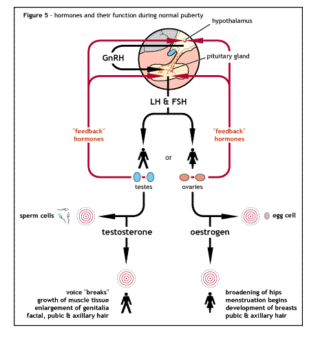
- The hypothalamus secretes gonadotrophin releasing hormone (GnRH) which travels in a small vein from the hypothalamus to the pituitary gland.
- GnRH stimulates in turn the anterior pituitary gland to secrete two hormones know as gonadotrophins. (A gonadotrophin is a hormone that stimulates a gonad, in this case the testis). These gonadotrophins are follicle stimulating hormone (FSH) and lutenizing hormone (LH). Also secreted in female they are glycoproteins.
- FSH acts by stimulating spermatogenesis by stimulating the sertoli cells to complete the development of spermatozoa from spermatids.
- LH stimulates the synthesis of the hormone testosterone by the leydig cells (interstitial cells) of the testis. It is therefore known interstitial cells stimulating hormone (ICSH) in the male
- Testosterone stimulates growth and development of the germinal epithelial cells (spermatogonia) to form sperms and also work with the FSH to stimulate the sertoli cells.
edu.uptymez.com
The negative feedback mechanism operates where by an increase in the level of testosterone results in a decrease in secretion of GnRH from the hypothalamus, this in turn results in declining levels of LH and FSH.
The testosterone also acts directly on the anterior pituitary gland to reduce LH secretion but this effect is weaker.
When the rate of spermatogenesis in high, inhibin (a glycoprotein hormone) is released, it acts on the anterior pituitary gland to reduce the secretion of FSH by negative feedback mechanism.
It also has a slight effect in the hypothalamus reducing GnRH secretion. When the rate of spermatogenesis low, inhibin is not secreted and FSH stimulates spermatogenesis.