THE ROLE OF CYCLIC AMP
Both FSH and LH acts by causing the release of cyclic AMP (Adenosine monophosphate) within the cells they stimulate.
Cyclic AMP is the second messenger system. It is released into the cytoplasm and then passes to the nucleus where it stimulates the synthesis of enzymes. In the case of LH, for example enzymes are
involved in the synthesis of testosterone from cholesterol.
STRUCTURE OF MATURE HUMAN SPERMATOZOANS
Structurally, a spermatozoan is divided into three pieces:-
I) Head piece
- This consists of a nucleus and small portion of cytoplasm.
- At the tip of the head, there is a special structure called acrosome. Acrosome is a collection of lysosomes; it thus contains very powerful hydrolytic enzymes known as proteases and hyaluronidases.
edu.uptymez.com
II) Middle piece
This is largely consisting of mitochondria. These provide energy for propelling the spermatozoans towards the egg cell. The head and middle peace together constitute the principal peace.
III) The tail piece
- It consists of the flagellum made of axial fillaments that continue from middle peace. The flagellum serves in:-
- Propelling the spermatozoans towards the egg cell.
- Orienting the spermatozoans so that it properly binds itself into the egg cell.
- At the end of the flagellum is a hair like extension called the end piece.
edu.uptymez.com
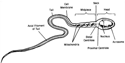
Structure of mature human sperm
ROLE OF SPERMATOZOAN.
- The role is to carry the paternal gamete materials into the egg cell so that after fertilization, the genetic makeup of the zygote is the mixture of the two maternal and paternal gamete materials.
edu.uptymez.com
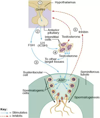
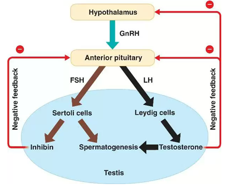
HORMONAL CONTROL OF SPERM PRODUCTION
- When the level of testosterone is low, the hypothalamus secretes a releasing hormone (called gonadotrophin releasing hormone or GnRH) in the blood.
- This peptide hormone flows in the blood directly to the pituitary a pre-sized organ hanging from the base of the brain where it stimulates the two peptide hormone, lutenising hormone (LH) and follicle stimulating hormone (FSH).
- These hormones (called gonadotrophins, since they stimulate gonalds) then move through blood stream and activate cells in the testis. LH triggers the intestinal cells to produce and secrete testosterone.
- FSH cause supporting cells (sertoli cells) to enhance formation of sperms.
- Soon the sperm count rise. Mean while, testosterone circulate in the blood stream at higher levels and the interconnected loop feeds back on itself. High testosterone levels signal the hypothalamus to produce less releasing hormone.
-
This inturn suppress the release of LH and FSH and without them less testosterone and fewer sperms are manufactured. In addition testosterone causes supporting cells in the testes to release the peptide hormone inhibin, which helps to inhibit FSH production.
When testosterone level drops too low again the hypothalamus is once more activated and the whole cycle starts again.
edu.uptymez.com
ADAPTATIONS OF THE SPERMATOZOANS.
The adaptations of the spermatozoans to its function include the following:-
- It has an acrosome that contains enzymes for digesting the egg cell membrane.
- It has numerous mitochondria that produce energy necessary for propelling the spermatozoans towards the egg cell.
- It has flagellum for propelling the spermatozoans for proper binding on the egg cells.
- Ability to sense the chemical attractants secreted egg cell so that its movement is directed toward source of chemicals.
- Ability to recognize and hence bind itself into the receptor sites on the surface of egg cells.
- Light nuclei and head piece following their changes, this enables it to move faster towards the egg cell.
edu.uptymez.com
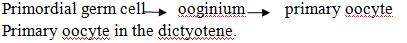
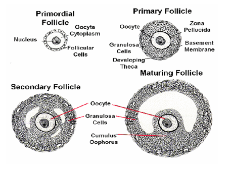
OOGENESIS
- Oogenesis begins soon after fertilization as primordial germ cell travel from the york sac to the gonalds, where they to proliferate mitotically.
- The gem cells multiply from only a few thousands to almost 7 million.
- They become oocytes once they enter the stages of meiosis several months after birth, now called primordial germ cells surrounded by fellicle cells from the somatic line. The oocytes are then arrested in the first meiotic phase until puberty.
- At puberty between 4 to 10 follicles begin to develop although only 1 – 2 are actually released.
- Surrounding each oocyote is a zona pellucida membrane granule and the cell layer.
- Each oocyte finishes its first meiotic division creating a secondary oocyte and polar body which serves no further functions.
- It begins the next meiotic cycle and is arrested in its second metaphase, at which point it is released from the ovary in ovulation.
- It will not finish the meiosis cycle until it encounters the stimuli of a sperm.
edu.uptymez.com
SUMMARY:
Oogenesis
At birth.
- Diploid cells in the ovary divide by mitosis from oogonia.
- Oogonia undergo meiosis I division to form primary oocyte steps at prophase I.
-
These remain in the follicles.
At puberty.
- Primary oocyte completes the 1st complete meiotic division to form polar bodies and secondary oocyte.
- Secondary oocytes undergo 2nd meiotic division and steps at metaphase II.
- Secondary oocyte is shed from ovary, if fertilized it complete its 2nd meiotic division to form ovum and polar bodies.
edu.uptymez.com
- Formation of the ovum involves substantial increase in cell volume as well as the acquisition of organelles that adapt the egg for reception of the sperm nucleus and support of the early embryo.
-
In the fetal ovary of mammals the oogonia undergoes meiotic divisions until the birth of the foetus, but the process involves the destruction of the majority of the developing ova by the seventh month of gestation reduces the number of oocytes from millions to a few hundred around the time of birth, the mitotic divisions ceases altogether and the fast female contains its full complement of potential ova.
edu.uptymez.com
| Weak of gestation | Stages | No of germ cells |
| ¾ | Primordial cells in the endoderm of the yolk sac. | |
| 5 – 6 | Premeiotic cells oogonia. | 10,000 |
| 8 | Propagation by mitosis. | 500,000 |
| 8 -20 | Mitosis, meiosis atresia maximum at week 20. | 6 – 700,000 |
| 20 -40 | Reduction of oocyte, 80% of germ cells are lost. | 1-2,000,000 |
| Birth to puberty | Further oocytes are lost by atresia. |
edu.uptymez.com
-
Unlike the formation of sperm in which the two divisions of meiosis produce four equivalent daughter cells, the cytoplasm of the oocyte is divided unequally so that three polar bodies with reduced
cytoplasm and one oocyte are final products.
-
Egg cytoplasm contains large stores of ribonucleic acid RNA in the form of ribosomal messenger and transfer RNA. These RNA’s direct the synthesis of proteins in the early embryo and have decisive
influence on the course of development.
edu.uptymez.com
DEVELOPMENT OF GERM CELLS IN THE OVARY
Following the immigration of the primordial germ cells into the gonadal ridge, they proliferate are enveloped by coelomic epithelial cells and form germinal cords that though keep their connection with the
coelom epithelium.
In the genital primodium, the following processes take place;
- A wave of proliferation begins that lasts from 15th weak to the 7th month.
edu.uptymez.com
Primary germ cells arise in the cortical zone via mitosis of oogonia dones, bound together in cellular bridges that happen in rapid succession.
The cell bridges are necessary for a synchronous onset of the subsequent meiosis.
- With the onset of meiosis earliest in the prophase in the 12th week the designation of the germ cells change. They are now called primary oocyte.
- The primary oocyte become arrested in the diplotene stage of prophase I the prophase of the 1st meiotic division.
-
Shortly before birth, all the total oocytes in female ovary have attained this stage.
The meiotic resting phase that then begins is called the dictyotene and it lasts till puberty during which each month and each month thereafter until menopause a pair of primary oocyte complete the first meiosis.
- Only few oocytes, secondary oocyte plus one polar body though reach the 2nd meiosis and the subsequent ovulation. The remaining oocytes that mature each month become atretic.
- The primary oocytes that remain in the ovaries stay in the dictyotene stages up to menopause. In extreme cases without ever maturing during the menstrual cycle.
- From birth, there are thus two different structures to be distinguished that at least conceptually do not develop further synchronously.
edu.uptymez.com
- Female germ called primary oocyte and which can develop further only during and after puberty hormonal cycle is necessary.
- Follicular epithelium, that can develop further from the primordial follicle via several follicle stages while oocytes remain in their primary states.
edu.uptymez.com
-
The developmental sequence of the female germ cell is as follows:-
The continous of the development/maturation of the oocyte begins again only a few days before ovulation.
- The developmental sequence of a follicle goes through various follicle stages.
edu.uptymez.com

- Since the follicle can die at any moment in this development (atresia) not all reach the tertiary follicle stage.
edu.uptymez.com
PRIMARY OOCYTE
- In the first week of the cycle the maturation of the oocyte in its associated follicle depends on the progress of maturation of the surrounding follicle cell.
- The fittest follicle with its oocyte becomes the dominant follicle in the second cycle week and later the graafian follicle.
- Up to just days before ovulation, the maturation of the oocyte consists in its ingestion of substances growth of the yolk, they are supplied by the granulose cells. This exchange of substances is mediated through cytoplast processes of the granulose cells that are anchored through zona pellucida at the ocoyte substance.
- The oocyte nucleus is also matured in the last days before the LH peak.
edu.uptymez.com
Up to this point it was arrested in the extremely elongated prophase (a dictyotene) of the first meiotic division, the corrested condition that has existed since the foetal period.
- Through the maturation the nucleus stages in the darkness of the prophase and prepares itself for the completion of the first meiosis which is triggered by the LH peak.
edu.uptymez.com
With the LH peak, the following maturation steps are now triggered in and around the oocyte up to ovulation.
In the oocyte:
- Termination of the first meiosis with ejection of the first polar body.
- Begin of the 2nd meiosis with arrest in the metaphase.
-
Maturation of the oocyte cytoplasm by preparing molecules and structures that will be needed at the time of fertilization.
In the follicle:
-
The granulosa cells that sit just outside on the zona pellucida withdraw their processes from oocyte surface back into the pellucida zona. These processes were in charge of transferring substances to
the oocyte.
- The periritelline space forms between the oocyte and the pellucida zona. This space is necessary for allowing division of the oocyte and for harbouring the first polar body formed in the division.
- Loosening of the granulosa cells in the vicinity of the cumulus oophorus and proliferation of the granulosa cells.
- Increasing the progesterone concentration in the follicle fluid via increased production in the granulosa cells.
edu.uptymez.com
Termination of the first meiosis
- The spindle apparatus for dividing the chromosomes has formed and oriented itself radically in the cellular surface.
- The first polar body will arise at the spot where the spindle apparatus is anchored on the cellular surface.
edu.uptymez.com
The process of the granulosa cells have refracted from the oocyte surface into the pellucida zona and this leads to the formation of the pervitelline space, in this space the ejection of the 1st polar body takes place as a sign that the 1st meiosis has ended.
- With the end of the first meiosis the name of the changes from primary oocyte to secondary oocyte.
edu.uptymez.com
The secondary oocyte
- Through the effect of LH on the granulosa cells, these have begun to loose their cellular bends and to multiply.
- They produce progesterone that is released into the fellicle fluid.
- Though the separation of the homologous chromosomes in the first meiosis a haploid (reduplicate) set of chromosomes is now to be found in the secondary oocyte.
edu.uptymez.com
The role of progesterone in the follicle fluid
Progesterone has the following two main tasks in the follicle fluid:-
- It stimulates the further maturation of the oocyte.
- During ovulation, it enters the fallopian tubes and guides the formation of a concentration gradient for attracting the sperm cells.
edu.uptymez.com
The follicle that is about to rapture:-
- Besides the hormones the granulosa cells also secrete an extra – cellular matrix, mainly hyaluronic acid, into the follicle fluid.
edu.uptymez.com
The cumulus cell bonds loosen further in this way together with the enclosed oocyte they free themselves from where they were attached to the follicular wall and in the follicle.
The wreath of granulosa cells that enclose the oocyte is called the corona radiate.
- The oocyte has now ended all the steps of maturation that were set into motion by the LH peak.
- The molecular and structural preparations for the time following the penetration by the sperm cell have now been made in the cytoplasm.
- A spindle apparatus (2nd meiosis) has again been able to form with the chromosome in the equational level (metaphase plate)
- The spindle is once more anchored radially to the cell membrane near the polar body.
edu.uptymez.com
The same processes of the spindle formation also take place in the polar body.
NB: The second meiosis is arrested in this position.
- Final steps of the maturation namely the freezing for the second meiosis are first completed by the second oocyte when the spermatozoa have penetrated the oocyte.
- The follicle and the oocyte are now ready for ovulation that takes place roughly 38 hrs after the LH peak.
edu.uptymez.com
ADAPTATION OF THE EGG CELL
- Has microvilli for nutrient absorption from follicular cells.
edu.uptymez.com
Follicular cells are the cells that usually surround the ovum when more layers are formed they tend to push away the follicular cells.
NB: The follicular cells are not part of the egg cell.
- It has stored food for zygote and embryo utilization (i.e. the yolk sac).
edu.uptymez.com
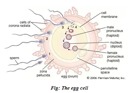
Fig: The egg cell
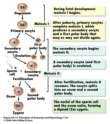
Fig: The stage of oogenesis
- Has cortical granules (Act as vesicles) that act to prevent polyspermy during fertilization.
edu.uptymez.com
This is done through two ways:-
- The process occurs very quickly and uses the same mechanism as in a chemical transmission of impulses. It occurs soon after the entry of a sperm cortical, are found in the cytoplasm.
edu.uptymez.com
They fuse with the membrane to release chemicals which haidens the membrane forming the fertilization membrane that prevents the entry of sperms.
-
Destruction of the sperm receptor sites. The sperms have sensors and ova have receptors-sperms move towards the ovum (like magnetic substance) chemotactically. The sperms receptor sites found
on the ovum are destroyed immediately after the entry of a sperm. This is done by the cortical granules.
2.Have receptor sites for spermatozoa to bind during fertilization.
3.Produce chemicals that attract sperms.
edu.uptymez.com
DIFFERENCES
| SPERMATOZOA | EGG CELL | |
| Small in size. | Larger than a sperm. | |
| Has a large nucleus. | Has a smaller nucleus. | |
| Has a very small amount of cytoplasm. | Has a very large amount of a cytoplasm. | |
| No food reserves (does not store food). | Stores large amount of food. | |
| Has acrosome. | Has no acrosome. | |
| No cortical granules. | Has cortical granules. | |
| Has head, middle piece principal and end piece. | No such division. | |
| No microvilli. | Has microvilli. | |
| Single layered. | Multi layered. | |
| It has flagellated and motile. | It lacks flagellum and non motile. | |
| It has numerous mitochondria. | Has few mitochondria. |
edu.uptymez.com
DIFFERENCE BETWEEN OOGENESIS AND SPERMATOGENESIS
| SPERMATOGENESIS | OOGENESIS | |
| Differentiation follow offer its meiotic division they are farmed only until the end of meiosis. | – Egg grows primarily in extend period of prophase i.e. prophase that is secondary oocyte is already matured. | |
| It occurs in male gonads i.e. testis. | – It occurs in female gonad i.e. ovaries. | |
| Four sperms are produced from one spermatogonium. | – Only one ovum is produced from one oogonium. | |
| The spermatocyte sperm mother cell divides by meiotic division into four equal sized cells and all the four cells are transformed into spermatozoa to act as reproductive unit. | -The oocyte divides unequally and produces a large sized ovum and three small sized bodies or polocytes which are sexually inert only ovum acts as reproductive unit. | |
| Spermatozoa are produced in large number. | -Ova are produced in large number. | |
| Spermatozoa are minute yolkless and motile. | – Ova are much large, often with yolk and non-motile. | |
| Continous production process although from puberty to old age sperm cells are being endangered, the production is subject to extreme fluctuations regalding both quality and quantity. | – Using up the oocyte generated before birth, continual decrease of the oocyte, beginning with the foetal period-exhaustion of supply at menopause. | |
| During foetal period no meiotic division, no germ cell production. | – During focal period, entering meiosis (arrested in dictyotene stage), there is a production of entire supply of germ cells. |